
Winning photo of 2020 Nikon Small World gives you a stunning close-up view of the zebrafish’s skeleton

© Daniel Castranova, Dr. Brant Weinstein & Bakary Samasa
Nikon Small World is one of those contests that shows us the world around us in a completely different light. The 2020 winners have been announced, and just like always, they reveal a stunning microscopic view of animals, plants, insects, and humans. We bring you the top 20 best photos from this year’s contest, and like every year, I’m sure you won’t be disappointed.
Now in its 46th year, the Nikon Small World competition received over 2,000 entries from scientists, artists, and hobbyists across 90 countries. I believe that the judges had a tough task of choosing the best. The panel of judges evaluated the photos on originality, informational content, technical proficiency, and visual impact.
The winners of this year’s contest are Daniel Castranova, Dr. Brant Weinstein, and Bakary Samasa from the USA. Their winning photo shows a dorsal view of the head of a zebrafish. The fluorescent bits represent the skeleton, the scales are shown in blue, and the lymphatic system in orange. Castranova stitched together more than 350 individual images to create this single photo. The image was acquired using a spinning disk confocal, merging together maximum intensity projections of three separate image Z stacks to generate the final reconstructed image.
1st Place
Daniel Castranova, Dr. Brant Weinstein & Bakary Samasa
Eunice Kennedy Shriver National Institute of Child Health and Human Development
National Institutes of Health
Section on Vertebrate Organogenesis
Bethesda, Maryland, USA
Dorsal view of bones and scales (blue) and lymphatic vessels (orange) in a juvenile zebrafish
Confocal
4X (Objective Lens Magnification)
What’s special about this photo isn’t only that it looks beautifully weird. It’s also very significant for the scientists because it shows that zebrafish have lymphatic vessels inside their skull. Before this discovery, this was believed to exist only in mammals. So what does this discovery mean? Well, it can lead scientists towards a revolution in research related to diseases that occur in the human brain, including cancer and Alzheimer’s. Naturally – this will also help in their treatment.
Daniel Knop from Germany was awarded the second place in the Nikon Small World competition. His image shows the embryonic development of a clownfish (Amphiprion percula) on days 1, 3 (morning and evening), 5, and 9, created using image-stacking.

2nd Place
Daniel Knop
Natur und Tier-Verlag NTV
Oberzent-Airlenbach, Hessen, Germany
Embryonic development of a clownfish (Amphiprion percula) on days 1, 3 (morning and evening), 5, and 9
Image Stacking
10X (Objective Lens Magnification)
Dr. Igor Siwanowicz won the third place for this picture of the tongue of a freshwater snail, using confocal microscopy. Look how beautiful and crazy this is!

3rd Place
Dr. Igor Siwanowicz
Howard Hughes Medical Institute (HHMI)
Janelia Research Campus
Ashburn, Virginia, USA
Tongue (radula) of a freshwater snail
Confocal
40X (Objective Lens Magnification)
In addition to the first three places, Nikon Small World recognized 88 more photos that stand out from over 2,000 entries. We bring you the top 20 in this article and take a look at the rest below. Make sure to visit the Nikon Small World’s website for more info, and if you’d like to see image collections from the previous years, here are some links:

4th Place
Dr. Vasileios Kokkoris, Dr. Franck Stefani & Dr. Nicolas Corradi
University of Ottawa & Agriculture and Agrifood Canada
Department of Biology
Ottawa, Ontario, Canada
Multi-nucleate spores and hyphae of a soil fungus (arbuscular mycorrhizal fungus)
Confocal
63X (Objective Lens Magnification)

5th Place
Ahmad Fauzan
Saipem
Jakarta, Indonesia
Bogong moth
Image Stacking
5X (Objective Lens Magnification)

6th Place
Dr. Robert Markus & Zsuzsa Markus
University of Nottingham
School of Life Sciences, Super Resolution Microscopy
Nottingham, Nottinghamshire, United Kingdom
Hebe plant anther with pollen
Confocal
10X (Objective Lens Magnification)

7th Place
Jason Kirk
Baylor College of Medicine
Optical Imaging & Vital Microscopy Core
Houston, Texas, USA
Microtubules (orange) inside a cell. Nucleus is shown in cyan.
Confocal
63X (Objective Lens Magnification)
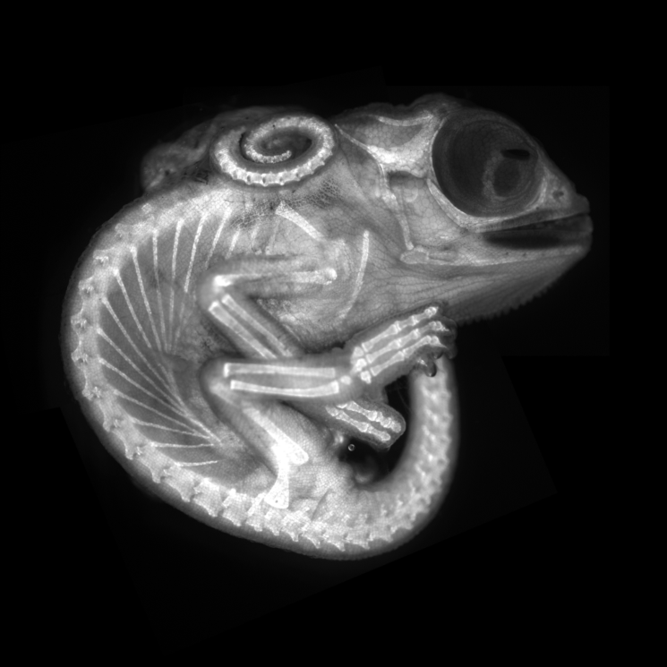
8th Place
Dr. Allan Carrillo-Baltodano & David Salamanca
Queen Mary University of London
School of Biological and Chemical Sciences
London, United Kingdom
Chameleon embryo (autofluorescence)
Fluorescence
10X (Objective Lens Magnification)

9th Place
Jason Kirk & Quynh Nguyen
Baylor College of Medicine
Optical Imaging & Vital Microscopy Core
Houston, Texas, USA
Connections between hippocampal neurons (brain cells)
Confocal
63X (Objective Lens Magnification)

10th Place
Ahmad Fauzan
Saipem
Jakarta, Indonesia
Daphnia magna (Phyllopoda)
Image Stacking
10X (Objective Lens Magnification)

11th Place
Dr. Tagide deCarvalho
University of Maryland, Baltimore County (UMBC)
Baltimore, Maryland, USA
Red algae
Confocal
63X (Objective Lens Magnification)

12th Place
Robert Vierthaler
Pfarrwerfen, Salzburg, Austria
Human hair
Image Stacking
20X (Objective Lens Magnification)

13th Place
Justin Zoll
Justin Zoll Photography
Ithaca, New York, USA
Crystals formed after heating an ethanol and water solution containing L-glutamine and beta-alanine
Polarized Light
4X (Objective Lens Magnification)

14th Place
Özgür Kerem Bulur
Istanbul, Turkey
Leaf roller weevil (Byctiscus betulae) lateral view
Image Stacking, Reflected Light
3.7X (Objective Lens Magnification)

15th Place
Dr. Eduardo Zattara & Dr. Alexa Bely
CONICET
Instituto Nac. de Investigaciones en Biodiversidad y Medio Ambiente
Bariloche, Rio Negro, Argentina
Chain of daughter individuals from the asexually reproducing annelid species Chaetogaster diaphanus
Brightfield
5X (Objective Lens Magnification)

16th Place
Alexander Klepnev
JSC Radiophysics
Moscow, Russian Federation
Nylon stockings
Polarized Light
9X (Objective Lens Magnification)

17th Place
Anne Algar
Hounslow, Middlesex, United Kingdom
Ventral view of an immature water boatman
Darkfield, Image Stacking, Polarized Light
4X (Objective Lens Magnification)

18th Place
Chris Perani
San Rafael, California, USA
Atlas moth wing
Image Stacking
10x (Objective Lens Magnification)

19th Place
Dr. Jan Michels
Christian-Albrechts-Universität zu Kiel
Department of Functional Morphology and Biomechanics
Kiel, Schleswig-Holstein, Germany
Silica cell wall of the marine diatom Arachnoidiscus sp.
Confocal
50x (Objective Lens Magnification)

20th Place
Dr. Dorit Hockman & Dr. Vanessa Chong-Morrison
University of Cape Town
Rondebosch, Cape Town, South Africa
Skeleton preparation of a short-tailed fruit bat embryo (Carollia perspicillata)
Brightfield
1X (Objective Lens Magnification)


